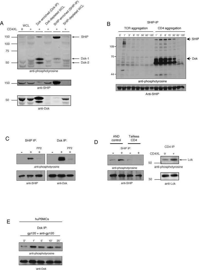
Figure 1. CD4 aggregation induces CD4 cytoplasmic tail-dependent tyrosine phosphorylation of SHIP and Dok.
(A) D10 cells were stimulated with anti-CD4 plus rabbit anti-rat IgG antibodies (2 min, 37°C) followed by lysis and immunoprecipitation using SHIP-, or Dok-specific antibodies directly conjugated to sepharose beads. Precipitates (5×10⁶ cell equivalents) and supernatants (1×10⁶ cell equivalents (WCL)) were resolved by SDS-PAGE, transferred to PVDF membrane, and analyzed by immunoblotting using phosphotyrosine-specific antibodies. (B) D10 cells were treated with biotinylated anti-TCR plus avidin, or anti-CD4 plus rabbit anti-rat IgG antibodies for the indicated time, followed by lysis and immunoprecipitation with SHIP-specific antibodies. Precipitates were resolved by SDS-PAGE, transferred to PVDF membrane, and analyzed by immunoblotting using phosphotyrosine-, or SHIP-specific antibodies. (C) D10 cells were pretreated with 1µM PP2, followed by anti-CD4 plus rabbit anti-rat IgG. Cells were lysed as described, and analyzed by immunoblotting using phosphotyrosine-, Dok-, and SHIP-specific antibodies. (D) Purified CD4+ T cells from AND.B10BR or tailless CD4 mice were stimulated with anti-CD4 plus rabbit anti-rat IgG. Cells were lysed and immunoprecipitated with SHIP-specific antibodies and analyzed as described above. (E) Freshly isolated huPBMCs were stimulated with gp120 plus goat anti-gp120 Ab for the indicated time, followed by lysis and immunoprecipitation with Dok-specific antibodies. Precipitates were resolved as described above, and analyzed by immunoblotting using phosphotyrosine- or Dok-specific antibodies. All results shown in figure 1 are representative of at least three independent experiments.

