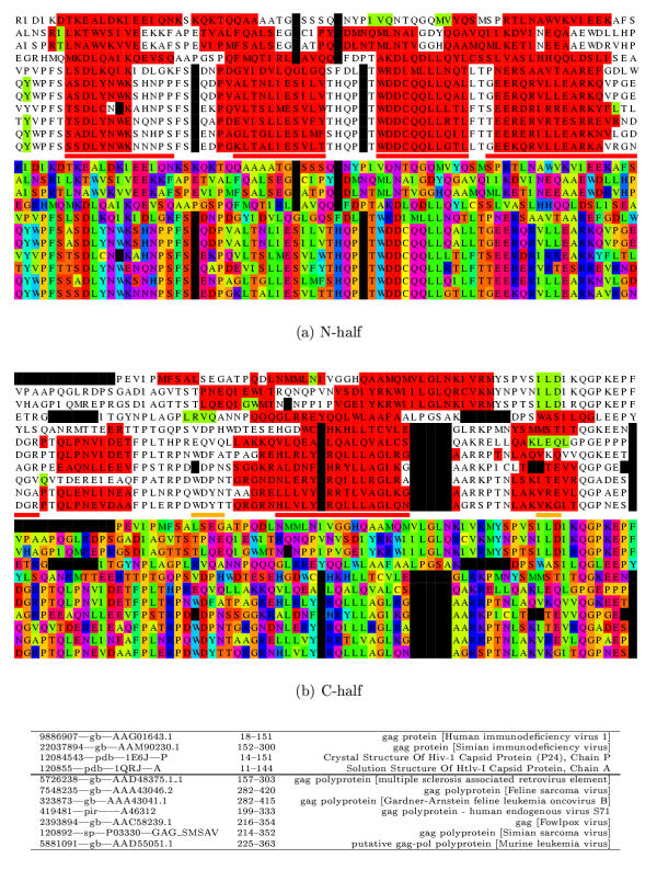
Figure 1.
MLV sequence alignment with the sequence of 1qrjA and related sequences. The alignment is displayed twice in different colouring schemes. In the top two panels, (a) the amino half is coloured firstly by predicted secondary structure (red = α, green = β) and then below using a different colour for each amino acid type (Taylor, 1997; black = gap). The known helices (N1–N5) marked as red lines (minor helices are orange) between the two blocks. In the lower two panels (b) the carboxy half of the alignment is shown in a similar way. The sequence identifiers are given below the alignment in the same order as they are aligned. The mid-line divides the 1qrjA sub-family from the MLV sub-family.

