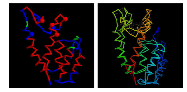
Figure 3.
Consensus model for the MLV N-domain. (a) The model for the MLV CA N-domain based on 1qrjA shown as a α-carbon trace with predicted α-helices coloured red (some fragments of predicted β-structure are coloured green). The molecule is in approximately the same orientation as in Figure 8(a) and residues identified by Stevens et al. (2003) are marked as small spheres. (b) The models based on 1qrjA and 1d1dA are shown superposed and coloured from blue (amino) to red (carboxy). Feint dashed lines connect identical residues. The wRMSd = 1.4 (uRMSd = 5.8) over 123 residues.

