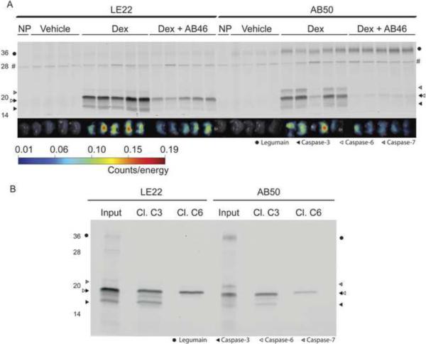
Figure 3. Comparison of LE22 and AB50 in dexamethasone-induced thymocyte apoptosis.
(A) Fluorescent images of apoptotic and control thymi and corresponding SDS-PAGE analysis. BALB/c mice were injected with dexamethasone or vehicle for 12 hours followed by injection of either LE22 or AB50 by tail vein. A subset of the dex-treated mice were pretreated with a caspase inhibitor, AB46, prior to probe injection. After four hours, thymi were harvested and imaged ex vivo for probe accumulation. Thymus proteins were then resolved by SDS-PAGE and scanned for Cy5 fluoresence using a flatbed scanner. An autofluorescent background protein in the no-probe control tumors is marked by an `#' (B) Immunoprecipitations of samples shown in (A). The first sample in each dex-treated lane for LE22 and AB50 was immunoprecipitated with the indicated antibody and analyzed by SDS-PAGE.

