Figure 2.
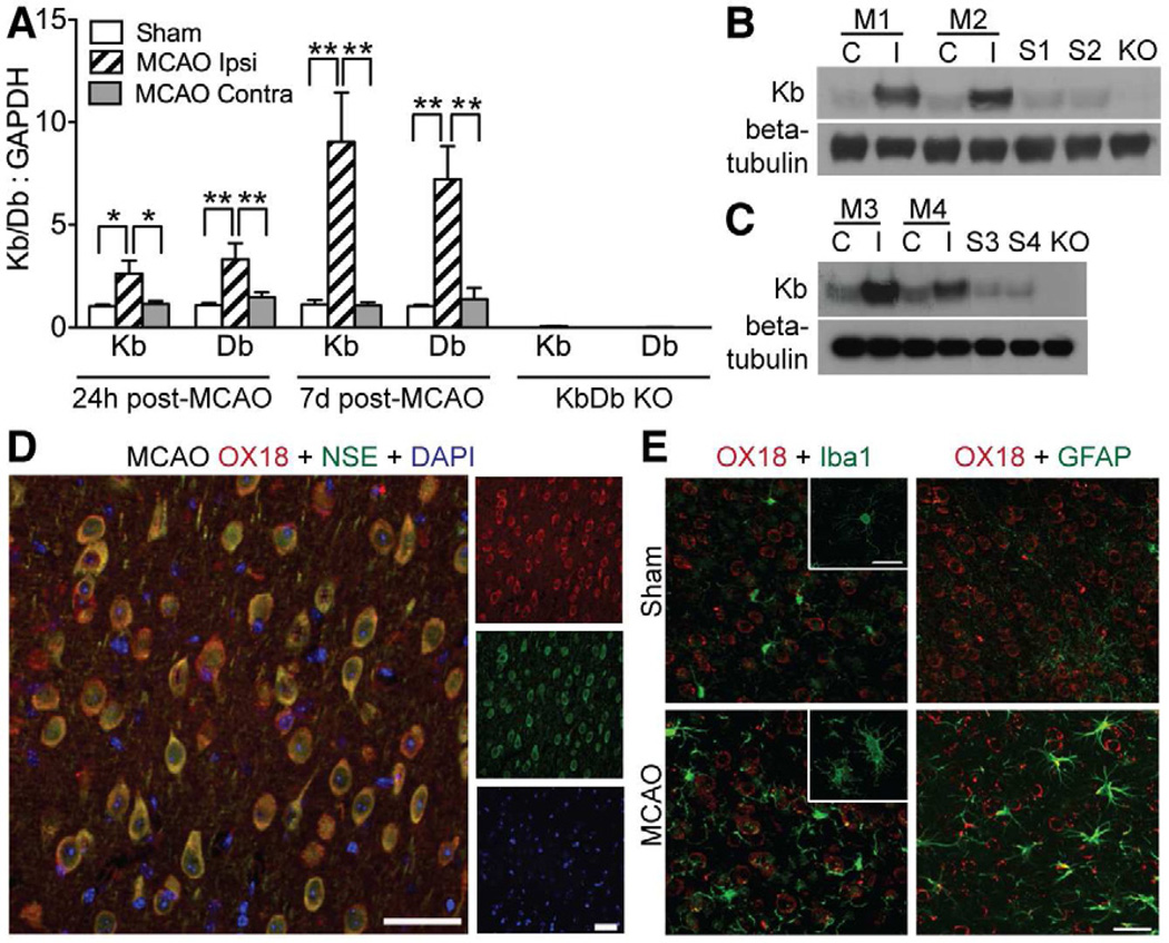
Expression of Kb and Db increases in brain of WT mice following MCAO. (A) Kb and Db gene expression was measured using qPCR at 24hr (n=7 for each experimental condition) or 7d (n=7 MCAO; n=4–8 Sham) post MCAO (Ipsi=Damaged hemisphere, Contra=undamaged hemisphere). Negative control data from KbDb KO samples demonstrates primer specificity (Kb:GAPDH=0.06 control relative to sham; Db:GAPDH=0.01 control relative to sham, p<10−8 for both). *P≤0.05, **p≤0.01. (B) Western blots showing protein expression of Kb 7d post MCAO in synaptosome-enriched preparations and (C) in synaptoneurosomes C=contralateral to injury; I=ipsilateral to injury; M1-4=different MCAO animals; S1-4=shams; KO=KbDb KO. (D, E) Immunohistochemistry of L5 in cortical penumbra using OX18 antibody. D: Majority of MHCI immunostaining (OX18; red) colocalizes with neuronal marker NSE (green) 7d post MCAO. E: GFAP (green; astrocytes) or Iba1 (green; microglia) 7d post MCAO. Scale bars D, E, 50μm. qPCR data represented as mean +/− SEM. See also Figure S2.

