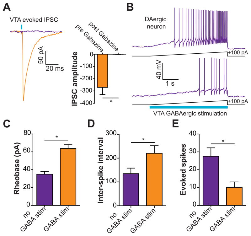
Figure 4. Activation of VTA GABA neurons reduces the excitability of VTA DA neurons and the release of DA in the NAc.
(A) Light evoked IPSCs recorded from VTA DA neurons are blocked by bath application of 10 μM Gabazine (t(4) = 4.5, p = 0.01, n = 5 neurons). Asterisk indicates p < 0.05. (B) Current clamp recordings in DA neurons in response to +100 pA current injection ramps paired with a 5 s optical stimulation of VTA GABA neurons. The top trace shows the evoked firing in a neuron induced by the current injection; bottom trace shows the response in the same neuron when neighboring VTA GABA neurons are coincidentally activated. (C) VTA GABA activation decreases the excitability of VTA DA neurons as indicated by an increase in rheobase (t(5) = 5.2, p = 0.004, n = 6 neurons). Asterisk indicates p < 0.05. (D,E) VTA GABA activation decreases the activity of DA neurons as indicated by an increase in the inter-spike interval (t(5) = 4.4, p = 0.007, n = 6 neurons) and a decrease in the total number of evoked spikes (t(5) = 6.7, p = 0.001, n = 6 neurons). Asterisk indicates p < 0.05.

