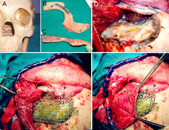
Figure 5.
(A) Titanium mesh blended using a standard skull model. (B and C) Superior orbital rim reconstructed with two calvarial split grafts and fixed to the orbitozygomatic bone. (D) Operative view after placing the titanium mesh and reattaching the orbitozygomatic complex. (E) Titanium mesh for the cranial defect. (F) Temporalis muscle sutured back in place. P, pericranial flap; T, temporalis muscle.

