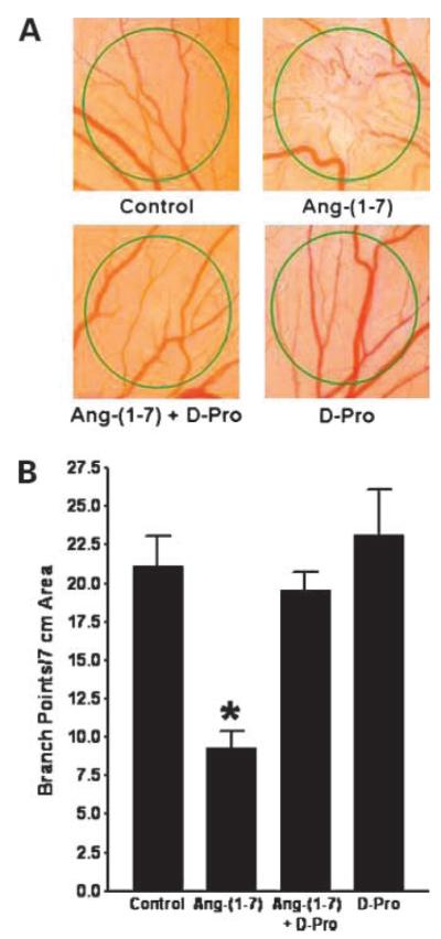
Figure 4.

Inhibition of neovascularization in the CAM by Ang-(1-7). Methylcellulose disks containing saline, 100 nmol/L Ang-(1-7), 100 nmol/L Ang-(1-7) and 1.0 μmol/L AT(1-7) receptor antagonist [d-Pro7]-Ang-(1-7) (D-Pro), or 1.0 μmol/L [d-Pro7]-Ang-(1-7) alone were placed in an avascular area of the CAM. After 24 h, the disks were photographed and branch points were quantified to assess vessel formation. A representative photograph of each treatment is shown in A and the quantification of branch points is depicted in B. *, P < 0.001; n = 9 to 12.
