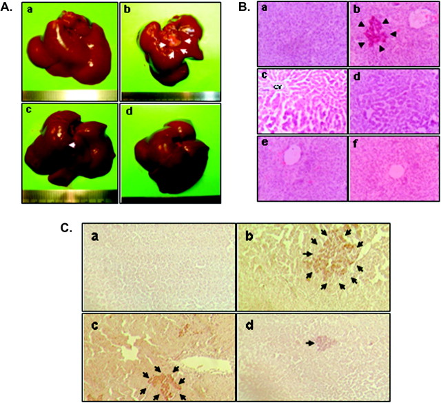
Fig. 2.
Pomegranate chemoprevention of DENA-initiated hepatocarcinogenesis in rats. (A) Morphological examination of rat liver tissue at the termination of the study. Macroscopically visible hepatic nodules are depicted by arrows. Representative livers were excised from several groups 22 weeks following the initiation of the study: (a) normal (group A) showing absence of nodules; (b) DENA control (group B) showing a large nodule; (c) PE at 1 g/kg body wt + DENA (group C) showing a small nodule and (d) PE at 10 g/kg body wt + DENA (group D) with no visible nodules. The animals fed PE at 10 g/kg only (group E) also showed no visible nodules (figure not shown). (B) Histopathological analysis of representative liver tissue from various experimental animals as observed by H&E staining (magnification: ×250). (a) Normal untreated rat liver (group A) showing normal cellular architecture; (b) DENA control (group B) showing eosinophilic foci and (c) areas of aberrant hepatocellular phenotype with irregular sinusoids and variation in nuclear size, shape and chromatin condensation; (d) liver section from PE (1 g/kg body wt) + DENA (group C) showing moderate improvement of hepatic histopathological indices over group B; (e) section from PE (10 g/kg body wt) + DENA (group D) showing hepatocytes exhibiting near-normal architecture and (f) section from PE (10 g/kg body wt) control group (group E) demonstrating characteristics of normal liver. CV, central vein. (C) Histochemical detection of hepatic GGT-positive foci in various rat groups (magnification: ×100). (a) Absence of foci in normal group; (b) a large GGT-positive focus in DENA control liver; (c) a medium size focus in low dose (1 g/kg) PE plus DENA-treated liver and (d) a small focus in high dose (10 g/kg) PE plus DENA-treated liver. GGT-positive foci of various sizes are indicated by arrows.

