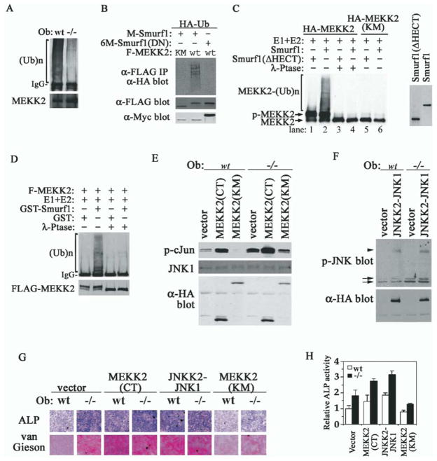
Figure 7. MEKK2 Is a Substrate of Smurf1-Mediated Ubiquitination and Activation of MEKK2-JNK Pathway Is Sufficient to Enhance Osteoblast Activity.
(A) Ubiquitination of endogenous MEKK2 in osteoblasts. Polyubiquitinated MEKK2 was detected by anti-ubiquitin Western analysis after precipitation of MEKK2 from cells that were pretreated with proteasome inhibitors.
(B) Smurf1 promotes MEKK2 ubiquitination. Ubiquitinated F-MEKK2 in Smurf1−/− MEFs was visualized by anti-HA-ubiquitin Western analysis after anti-FLAG immunoprecipitation. Expression of Myc-tagged Smurfs is shown in bottom panel.
(C) In vitro polyubiquitination of MEKK2 by Smurf1 detected by anti-HA blot. HA-MEKK2 and HA-MEKK2(KM) were isolated by anti-HA antibody from transfected Hep3B cells. Myc-tagged Smurf1 and Smurf1(ΔHECT) were in vitro translated and are shown in the right panel.
(D) In vitro ubiquitination of MEKK2 by purified GST-Smurf1 detected by anti-ubiquitin blot. FLAG-MEKK2 purified from transfected Drosophila S2 cells is shown at bottom panel.
(E) In vitro kinase assay of JNK1 after immunoprecipitation from osteoblasts expressing MEKK2 mutants. GST-c-Jun was used as the kinase substrate and was analyzed by anti-p-c-Jun blot. Total JNK1, HA-MEKK2(CT), and MEKK2(KM) are shown below.
(F) Western analyses of phosphorylated JNK in osteoblasts expressing HA-JNKK2-JNK1, which does not affect endogenous JNK activity. Arrows, endogenous p-JNK1/2; arrowhead, p-JNKK2-JNK1 fusion.
(G) Ectopic expression of activated MEKK2 and JNK1 is sufficient to enhance osteoblast activity. Staining for ALP activity and collagen matrix was carried out after incubating in differentiation medium for 16 days.
(H) ALP quantification of cells in (G) after differentiation for 12 days.

