FIG. 5.
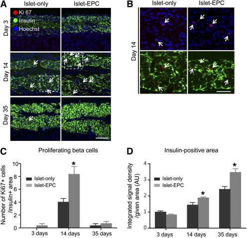
EPC cotransplantation is associated with β-cell proliferation. A–C: The transplanted islets were evaluated for proliferation by Ki67/insulin double staining at days 3, 14, and 35. A: The Ki67/insulin–double-positive proliferating β cells were visualized at each time point (arrow). Hoechst stain was used to distinguish the nucleus. B: Magnification views of the inserts within the white dotted line at day 14. C: The number of Ki67-positive cells among the insulin-positive cells in the total given area was counted (0.18 mm2) (n = 4). D: The integrated density of the insulin-positive area of the total given area (0.18 mm2) was measured and presented in arbitrary units (AU), with the value of day 3 for the islet-only group set to 1 (n = 4). The data are presented as means ± SE. *P < 0.05 vs. the islet-only group. Scale bars, 100 μm in A and 50 μm in B. (A high-quality color representation of this figure is available in the online issue.)

