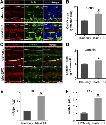
FIG. 6.
β-Cell proliferation in the EPC-cotransplanted group is associated with more basement protein and HGF production. A–D: At day 14, the BS1-lectin–positive blood vessels and Col IV– or laminin-positive basement membrane were visualized. The insulin was immunostained to distinguish the graft site. The representative images for Col IV (A) and laminin (C) are shown. White dotted lines show the territory of the graft site. Scale bars, 100 μm. The Col IV–positive (B) and the laminin-positive (D) areas from total graft area were calculated as a percentage of the total graft area and then presented as arbitrary units (AU), with the value of the islet-only group set to 1 (n = 4). E and F: The graft sites of day 10 from each mouse in vivo (E) and human EPCs cocultured in vitro with or without porcine islets (F) were harvested. The specimens were analyzed for HGF by q-PCR using primers that functioned for all three species (human, porcine, and mouse) in E and for human exclusively in F. The data are presented in arbitrary units after normalization to glyceraldehyde-3-phosphate dehydrogenase, with the value for the islet-only group set to 1 in E and with the value for the EPC-only group set to 1 in F (n = 4). The data are presented as means ± SE. *P < 0.05 vs. the islet-only group in B, D, and E and vs. the EPC-only group in F. (A high-quality color representation of this figure is available in the online issue.)

