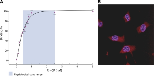
FIG. 1.
A: Binding of rhodamine (Rh)-labeled C-peptide (CP) to membranes of human renal tubular cells. Fractional saturation of the membrane-bound ligand is presented as a function of the C-peptide concentration in the surrounding medium. The light blue area represents the physiological concentration (conc) range. Data are from Ref. 13. B: Intracellular distribution of Rh-labeled C-peptide examined with confocal microscopy. Swiss 3T3 fibroblasts were incubated with Rh-labeled C-peptide (1 μmol/L) for 30 min at 37°C. Overlay graph of nuclear staining with Hoechst dye 33342 (1 μg/mL, blue) and Rh-labeled C-peptide staining (red). Treatment with Rh alone did not result in cytosolic or nuclear staining. Reprinted with permission from Lindahl et al. (19). (A high-quality digital representation of this figure is available in the online issue.)

