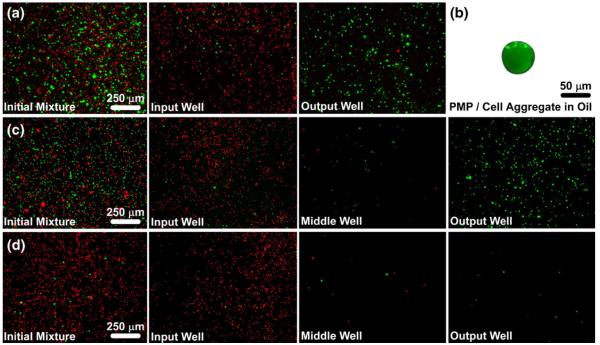
Fig. 2.
Selective isolation of green fluorescent MCF-7 cells from red fluorescent particles. (a) A mixture of cells (green), particles (red) and anti-EpCAM-labeled PMPs were added to an IFAST device, which was used to transfer the cells to the output well of the device while leaving most of the particles in the input well. (b) The PMPs and cells form a tight aggregate as they cross the oil phase, thus minimizing carryover of extraneous material, such as the particles. (c) The same sample from A was purified using an IFAST device with two oil barriers in series with a washing buffer located between them. The double oil barrier increases purity since nearly all of the contaminating particles that cross the first oil barrier do not cross the second oil barrier. (d) An additional IFAST with two oil barriers was loaded with a mixture containing approximately 10× fewer cells than in A and B. The double oil barrier is suitable for isolating small cell numbers with minimal contamination. A, C, and D were taken at 4× magnification while B was taken at 20× magnification. Images of input, middle, and output wells were taken after IFAST purification

