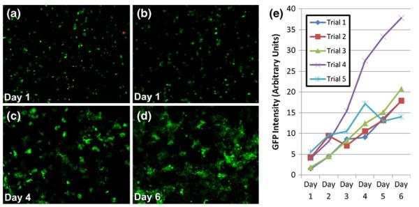
Fig. 5.
Viability of GFP-positive MCF-7 cells following traverse through oil barrier. (a) Cells seeded into 96-well plate following oil traverse and stained to highlight dead cells (orange). (b-d) Proliferation of MCF-7 cells in 96-well plate following traverse through the oil barrier. Images taken at 1, 4, and 6 days after traverse, respectively. (e) Quantification of GFP signal for five independent trials where MCF-7 cells were drawn across oil barrier and seeded into wells of a 96-well plate. Note that GFP intensity is proportional to cell number (Fig. S-1)

