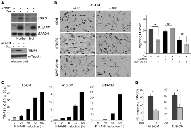
Figure 3. Induction of TIMP3 expression by P14ARF inhibits EC migration.
(A) Northern blot (upper panel) and Western blot (lower panel) demonstrating the silencing of TIMP3 with siRNA in A5 glioma cells. (B) Modified Boyden chamber migration assay showing that TIMP3 is required for the inhibition of EC migration by the CM of P14ARF-expressing cells (2 μg/ml dox for 48 hours). Prior to CM collection, the A5 cells were transfected with negative control or TIMP3 siRNAs. HDMECs were seeded in the upper chamber with medium containing 1× CM (diluted from 30× concentrated), and the migration across gelatin type B–coated filters was performed. The migrated cells were photographed (left panel) and counted (right panel). The MMP-2/9 inhibitor I (inh) was used at 650 nM. Scale bar: 200 μm. (C) ELISA quantification of TIMP3 production in serum-free culture medium of A5, A18, and C19 cells with or without P14ARF induction by dox for up to 5 days. Data are expressed in ng/100 μl of CM and as mean ± SD. Each experiment was performed in duplicate. (D) Modified Boyden chamber migration assay showing the effect of 5-day CM of A18 and C19 cells with or without dox on EC (HDMEC) migration. The assay was performed as described in B. The unpaired 2-tailed Student’s t test was used for B and D to assess the statistical differences between experimental groups, *P <0.05; n = 3.

