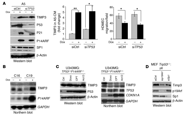
Figure 4. The upregulation of TIMP3 gene expression by P14ARF is independent of P53.
(A) Effect of TP53 silencing on the induction of TIMP3 by P14ARF and on EC migration. Left panel: Western blot showing that the silencing of TP53 expression by siRNA in A5 cells does not affect the induction of TIMP3 expression by P14ARF. Note that the silencing of TP53 has no effect on SP1 protein levels. Middle panel: ELISA quantification of TIMP3 levels in CM of A5 cells previously transfected with either negative control siRNA (siCtrl) or TP53 siRNAs (siTP53) and cultured in medium with or without dox without serum for 48 hours. The assay was carried out in triplicate, and results are expressed as mean ± SD. Right panel: Boyden chamber HDMEC migration assay in the presence of CM from control and TP53 siRNA–transfected A5 cells with or without dox. Results are expressed as mean (±SD) cell number per field. *P < 0.05, **P < 0.01, unpaired 2-tailed Student’s t test. (B) Northern blot showing that P14ARF strongly activates TIMP3 mRNA expression in TP53-null C19 cells. (C) Western blot (left) and Northern blot (right) showing that activation of endogenous WT P53 with genotoxic agents does not induce TIMP3 protein or mRNA expression. U343MG cells were treated for 24 hours with doxorubicin (Doxo; 1 μg/ml), camptothecin (CPT; 5 μM), or actinomycin D (Act D; 10 nM) to activate P53. Note that P53 activation by genotoxic agents strongly increases its stability, yet TIMP3 protein levels remain unchanged. (D) Western blot analysis showing that silencing p19Arf or Sp1 in Trp53-null MEFs (passage 8) decreases Timp3 expression.

