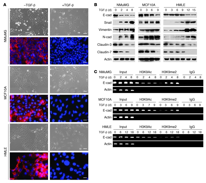
Figure 1. H3K9 methylation at the E-cadherin promoter is associated with TGF-β–induced EMT in three model cell lines.
(A) NMuMG, MCF10A, and HMLE cells were treated with TGF-β1 (5 ng/ml) for 3, 9, and 12 days, respectively; cell morphological changes associated with EMT are shown in the phase contrast images. Expression of E-cadherin (red) was analyzed by immunofluorescence staining. Nuclei were visualized with DAPI staining (blue). Scale bars: 50 μm. (B) NMuMG, MCF10A, and HMLE cells were treated with TGF-β1 (5 ng/ml) for the indicated time periods, and expression of E-cadherin (E-cad), claudin-3, claudin-7, Snail, N-cadherin, and vimentin in these cells was analyzed by Western blotting. (C) NMuMG, MCF10A, and HMLE cells were treated with TGF-β1 (5 ng/ml) for different time periods, and H3K9me2 and H3K9Ac at the E-cadherin promoter in these cell lines were analyzed by ChIP assay.

