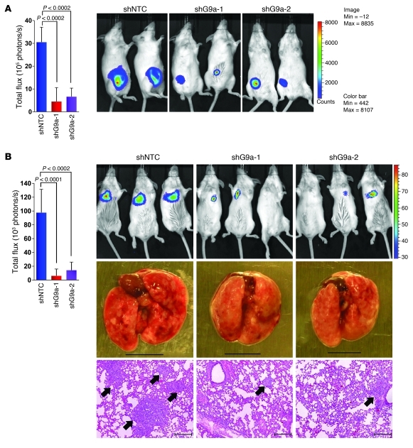
Figure 9. Knockdown of G9a expression suppresses breast tumor growth and lung colonization in vivo.
(A) MDA-MB231 cells stably expressing control vector or G9a shRNA were injected into the mammary fat pad of ICR-SCID mice. The growth of breast tumors was monitored every 3 days. After 9 weeks, the size of tumors from each group was recorded by using bioluminescence imaging and quantified by measuring photon flux. Values are the mean of 6 animals ± SEM. (B) Cells from A were also injected into the tail vein of ICR-SCID mice. After 9 weeks, the development of lung metastases was recorded using bioluminescence imaging and quantified by measuring photon flux (mean of 6 animals ± SEM). Results for 3 representative mice from each group are shown. Mice were sacrificed, and lung metastatic nodules were examined macroscopically or detected in paraffin-embedded sections stained with H&E. Scale bars: 100 μm. Arrowheads indicate lung metastases.

