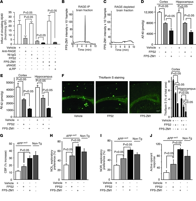
Figure 3. FPS-ZM1 and FPS2 block RAGE-mediated Aβ BBB transport, Aβ pathology, and functional outcome in old APPsw/0 mice.
(A) Influx of circulating 125I-Aβ40 (1.5 nM) across the BBB in 15- to 17-month-old APPsw/0 mice with and without anti-RAGE antibody (40 μg/ml), NI IgG (40 μg/ml), FPS2 (200 nM), or FPS-ZM1 (200 nM) alone or after preincubation with sRAGE or sLRP. Values are mean ± SEM; n = 4–6 mice per group. (B and C) Detection of FPS-ZM1 in the brain of 15 month old APPsw/0 mice 5 minutes after an I.V. administration. FPS-ZM1 was found in RAGE-IP brain fraction (B), but not in RAGE-depleted brain (C). Representative results from 6 experiments are shown. (D and E) Aβ40 (D) and Aβ42 (E) levels in the cortex and hippocampus. (F) Thioflavin S–positive amyloid deposits in the brain (left) and quantification of thioflavin S–positive area in the cortex and hippocampus (right). Scale bar: 350 μm. (G–J) CBF response to whisker stimulation (G), NOL (H), NOR (I), and active operant response (J) in 15-month-old APPsw/0 mice treated with vehicle, FPS2, or FPS-ZM1 for 2 months. Nontransgenic littermate controls (gray columns) are shown in panels G–J. Values are mean ± SEM. n = 4–6 mice per group.

