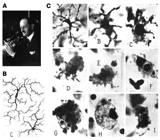
Figure 3. Microglial cells, as described by Pio del Rio-Hortega (161).
(A) Pio del Rio-Hortega (1882–1945), who characterized and named microglial cells. (B) Images of ramified microglial cells drawn by Hortega. (C) Morphological transformation of microglia to phagocytic macrophage. Panels as lettered in C: A, Microglial cell with modestly thickened processes as compared with ramified microglia; B, microglial cell with short, thick processes and enlarged cell body; C, microglial cell with pseudopodia; D, amoeboid microglial cell; E, amoeboid microglial cell with pseudopodia; F, microglial cell with phagocytosed leukocyte; G, microglial cell with numerous phagocytosed erythrocytes; H, microglial cell with lipid inclusions, also termed “foam cell”; I, microglial cell in mitotic division. Reproduced with permission from Physiological Reviews (161).

