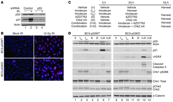
Figure 7. p53 status is a key determinant of how TNBCs respond to DNA damage followed by Chk1 inhibition.
WU-BC3 cells were infected with control retroviruses or retroviruses encoding p53-specific shRNAs to generate the BC3-p53WT and BC3-p53KD lines, respectively. (A and B) Cells were either mock irradiated (–) or exposed to 10 Gy IR (+) and then monitored for the integrity of the p53 pathway by observing changes in the levels of p53 and p21 by Western blotting (A) and for the integrity of HRR by monitoring for the appearance of Rad51 foci (B; pink stain, Rad 51). Original magnification, ×400. (C) Timeline of treatment of cells with either vehicle, irinotecan, AZD7762, the Chk2 inhibitor, or irinotecan in combination with AZD7762 or the Chk2 inhibitor. Cell lysates were prepared and analyzed for the indicated proteins by Western blotting (D).

