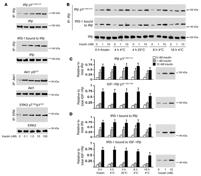
Figure 1. Ex vivo stimulation is a valid method for studying insulin signaling in postmortem HF.
(A) Representative dose-response tests in 1 of 5 N adult humans at 0–100 nM insulin in immunoblots of phosphorylated or bound antigens from immunoprecipitates of the indicated antigens. (B) Sample blots on the rat HF, showing that PMIs up to 16 hours (n = 4 per PMI) had no substantial effect on basal levels of IRβ or signaling evoked by 1 or 10 nM insulin. Effects of insulin on IGF-1 signaling were also tested on the same samples (Supplemental Figure 2). (C) PMI effects on IRβ pY and IGF-1Rβ pY relative to total receptor levels (mean ± SEM). 1 nM insulin activated IRβ (P = 0.0015), but not IGF-1Rβ (see blots). 10 nM insulin induced greater activation of IRβ (P = 0.0063) as well as IGF-1Rβ (P = 0.0009). (D) PMI effects on IRS-1 bound to IRβ and IGF-1Rβ relative to total receptor levels (mean ± SEM). 1 nM insulin induced IRβ binding (P = 0.0002), but not IGF-1Rβ binding (see blots), of IRS-1. 10 nM insulin induced greater IRβ binding (P = 0.0009) as well as IGF-1Rβ binding (P = 0.0007) of IRS-1. kDa values correspond to the molecular weight marker closest to the bands shown. *P < 0.005.

