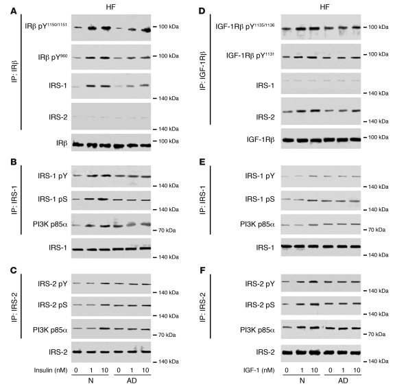
Figure 4. Ex vivo stimulation revealed IRS-1–associated insulin resistance and IRS-2–associated IGF-1 resistance in HF of AD cases.
(A–C) Western blots from a representative matched pair of N and AD cases showed decreased signaling responses in the AD case to 1 and 10 nM insulin without affecting IRS-2 (specifically, reductions in IRβ activation; IRS-1 binding of IRβ, IRS-1 activation [pY] and suppression [pS]; and PI3K p85α binding of IRS-1). (D–F) Western blots from a representative matched pair of N and AD cases showed decreased signaling responses in the AD case to 1 and 10 nM IGF-1 without affecting IRS-1 (specifically, reductions in IGF-1Rβ activation; IRS-2 binding of IGF-1Rβ, IRS-2 activation and suppression; and PI3K p85α binding of IRS-2). See Figure 5 and Tables 1–4 for quantification.

