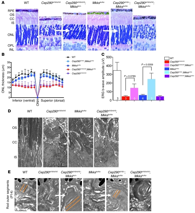
Figure 4. Triallelic loss of Mkks and/or the Cep290-DSD domain ameliorate cilia phenotypes in photoreceptors.
(A) Cross sections through the P18 retina in different combinations of Cep290rd16 and Mkksko alleles, as indicated. Note the short, abnormal OSs in Cep290rd16/rd16 or Mkksko/ko genotypes and the more normal OS in the triallelic Cep290rd16/+;Mkksko/ko genotype. Here, the Cep290rd16/rd16;Mkksko/ko genotype looks similar to Cep290rd16/rd16. The white arrows indicate comparison between 2 similar genotypes that are improved by combining alleles of Cep290 and Mkks. Original magnification, ×40. (B) Quantitation of outer nuclear layer thickness at P18 in the genotypes indicated. Higher variability is noted in double-homozygous mutants (see error bars on Cep290rd16/rd16 versus Cep290rd16/rd16;Mkksko/ko). Error bars are SD; n = 6 (WT), n = 4 (Cep290rd16/+;Mkksko/ko), n = 3 (Mkksko/ko), n = 14 (Cep290rd16/rd16;Mkksko/ko), n = 8 (Cep290rd16/rd16). (C) Scotopic ERG b-wave amplitudes in the indicated mouse genotypes (at P20). Removing one WT Mkks allele on a Cep290rd16/rd16 background results in improved responses, as does adding one Cep290rd16 allele on a Mkksko/ko background. Note that single homozygous or double-homozygous genotypes have essentially no ERG b-wave response. Error bars are SD; n = 3 (WT), n = 3 (Cep290rd16/rd16), n = 6 (Cep290rd16/rd16;Mkksko/+), n = 4 (Mkksko/ko), n = 3 (Cep290rd16/+;Mkksko/ko), and n = 3 (Cep290rd16/rd16;Mkksko/ko). (D) Longitudinal EM sections through the OS, connecting cilia, and inner segments in P14 retina show that OS morphology is disrupted in the indicated mutant genotypes. Original magnification, ×3,000. (E) Higher-magnification images (original magnification, ×30,000) of OSs in P14 retina confirm improved OS morphology in Cep290rd16/+;Mkksko/ko and Cep290rd16/rd16;Mkksko/+ triallelic genotypes. OSs of triallelic mice form concentric stacks of discs (parallel orange lines), more similar to WT. The white arrows indicate comparison between 2 similar genotypes that are improved by combining alleles of Cep290 and Mkks. RPE, retinal pigment epithelium; CC, connecting cilia; IS, inner segment; ONL, outer nuclear layer; OPL, outer plexiform layer; INL, inner nuclear layer; ONH, optic nerve head.

