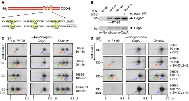
Figure 1. Analysis of CagAPY protein species during infection with H. pylori by 1-DE and 2-DE.
(A) CagA proteins vary in the C-terminal EPIYA phosphorylation sites depending on geographical origin. Western CagA proteins (e.g., strain 26695) contain the EPIYA-A, EPIYA-B, and EPIYA-C segments, while East Asian strains contain the EPIYA-A, EPIYA-B, and EPIYA-D segments (e.g., strain TN2-GF4). These motifs are tyrosine phosphorylation sites that can be phosphorylated by Abl and Src tyrosine kinases (10–13). (B) AGS cells were infected for the indicated times with strain 26695. The resulting protein lysates were separated by 1-DE, and phosphorylation of injected CagA was examined using α–PY-99 and α-CagA antibodies (arrows). (C) Separation of CagA protein species from B by 2-DE. Depending on the time of infection, full-length CagAPY appeared as 1 spot (spot 1, red arrows, pI = 7.0) or 2 spots (spots 1 and 2; spot 2, green arrows, pI = 6.5) as indicated. The α-CagA antibody probe revealed a third spot (spot 3, blue arrows, pI = 7.5). Overlay of both exposures yielded 2 or 3 spots as shown. Strain TN2-GF4 exhibited the same pattern as 26695 (bottom). (D) Inhibition of Src with PP2 (10 μM) or Abl with SKI-DV2-43 (1 μM) revealed significant changes in spot intensity depending on the time of infection.

