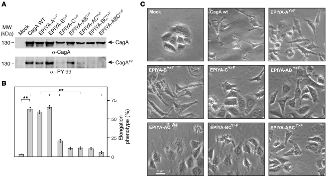
Figure 4. Role of EPIYA motifs in CagA phosphorylation and AGS cell elongation during H. pylori infection.
(A) AGS cells were infected for 4 hours with CagA-expressing H. pylori strains as indicated. Phosphorylation of CagA was examined using α–PY-99 and α-CagA antibodies (arrows). (B) The number of elongated cells in each experiment was quantitated in triplicate in 10 different 0.25-mm2 fields. (C) Phase-contrast micrographs of AGS cells infected with the different strains as indicated. **P ≤ 0.001.

