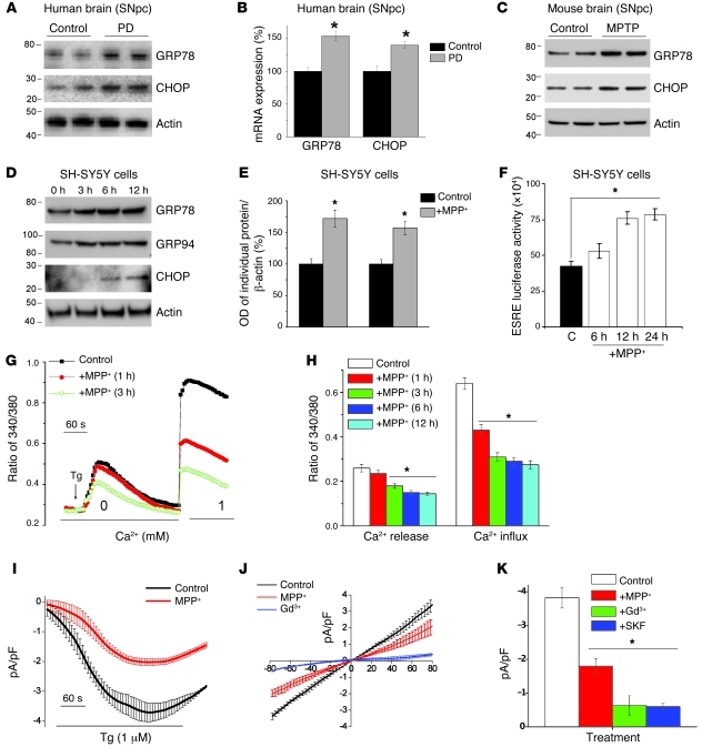
Figure 1. MPP+ induces ER Ca2+ depletion by attenuating SOCE, which causes ER stress.
(A) Representative blots from the SNpc region of postmortem human PD (n = 5) and age-matched controls (n = 4). (B) RNA was extracted, and RT-PCR was performed. Values represent mean ± SD from 3 independent experiments (*P < 0.05). (C) Mice received 25 mg/kg MPTP as described in ref. 19, and SNpc samples were removed, processed, and immunoblotted using the respective antibodies. (D) SH-SY5Y cells were treated with 500 μM MPP+, and cell lysates were resolved and analyzed by Western blotting. Antibodies are labeled; β-actin was used as loading control. (E) Quantification (mean ± SD) from 3 or more independent experiments. The OD of GRP78 and GRP94 was normalized to β-actin. (F) Cells transfected with the ERSE promoter were lysed after MPP+ treatment, and luciferase assays were performed. Values represent mean ± SD from 3 independent experiments (*P < 0.05). (G) Analog plots of the fluorescence ratio (340/380 nm) from an average of 30–40 cells in each condition. (H) Quantification (mean ± SEM) of 340/380 ratio. *P < 0.05 versus untreated. (I) Tg-induced currents (mean ± SEM) were evaluated in control and MPP+-treated (12 hours) cells. The holding potential for current recordings was –80 mV. (J) I-V curves (mean current ± SEM) under these conditions; the average (8–10 recordings) current intensity under various conditions is shown in K. *P < 0.05 compared with control; values are shown as mean ± SEM. SKF, SKF-96365.

