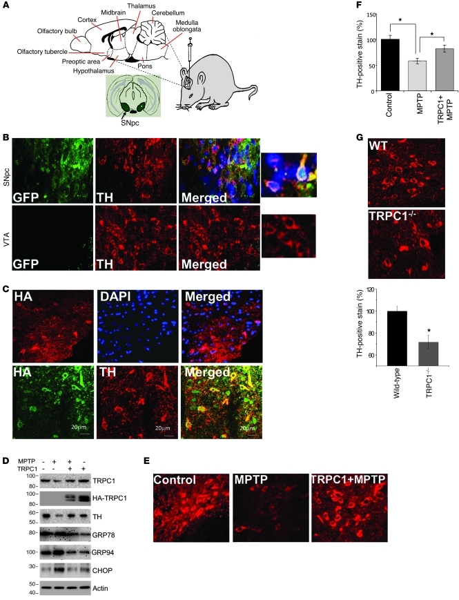
Figure 6. TRPC1 overexpression protects DA neuron in an in vivo MPTP model of PD.
(A) Graphic representation of intranigral injection protocol. (B and C) Unilateral injection of GFP or HA-TRPC1 adenovirus (3 × 10⁷ particles) into the SNpc (n = 6). Brain samples containing the SNpc region were sectioned (12 μm) and stained for TH immunofluorescence. Expression of GFP and TH in the SNpc and in ventral tegmental area (VTA) were evaluated. HA-TRPC1 colocalized with TH (C, bottom row). Scale bars in C: 20 μm. (D) HA-TRPC1 or GFP adenovirus was injected directly into the SNpc of animals (n = 6–8 per group) 7 days before MPTP treatment. After 1 week of MPTP injection, SNpc samples were removed and subjected to SDS-PAGE and immunoblotted with the respective antibodies. Control GFP virus–injected mice received an equal volume of saline. (E) Representative TH staining of the ipsilateral sides of animals injected with the indicated virus with and without MPTP. (F) Quantification of the number of TH-positive neurons from ipsilateral or contralateral sides for the indicated treatment groups. Data are mean ± SEM. *P < 0.05. (G) Brain samples containing the SNpc from wild-type and Trpc1–/– mice were sectioned (12 μm) and stained for TH. Quantification of TH-positive neurons is shown in the graph. Data are presented as mean ± SEM. *P < 0.05. Original magnification, ×40; magnified images in B, ×100.

