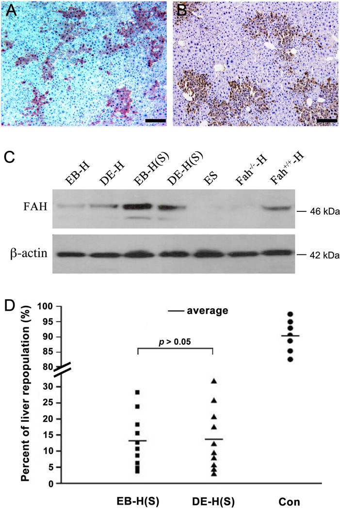
Figure 3. Serial transplantation of ES cells derived hepatocytes.
(A, B) FAH positive hepatocytic nodules in liver samples of secondary recipients after serial transplantation of ES cells derived hepatocytes under EB (A) or DE methods (B). (C) Western blot of liver samples showed the levels of FAH protein expressed in the repopulating hepatocytes after primary and secondary transplantations. (EB-H represents the repopulating hepatocytes derived under EB method; DE-H represents the repopulating hepatocytes derived under DE method; EB-H (S) or DE-H (S) represent either of the repopulating hepatocytes after serial transplantation; ES represents the liver sample of recipients with transplantation of ES cells without differentiation; Fah−/−-H represents liver sample of Fah−/− mice without cell transplantation; Fah+/+-H represents liver sample of 129S4 wild-type mice. (D) Comparison of liver repopulation levels of ES cells derived hepatocytes under EB or DE methods in serial transplantation (Liver repopulations transplanted with wild-type hepatocytes were used as control). Scale bars: 200 µm (A and B).

