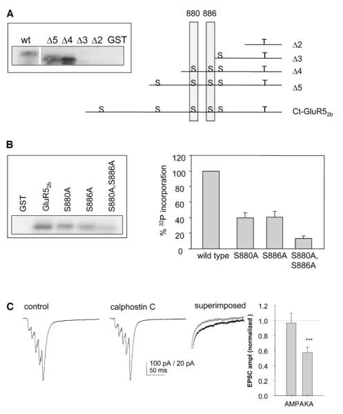
Figure 7.
PKC Phosphorylates GluR52b and Inhibition of PKC Activity Reduces KAR-Mediated EPSCs
(A) Schematic of the truncation mutants generated and consensus PKC phosphorylation sites (S) on ct-GluR52b. The insert panel shows in vitro phosphorylation of each of the truncations by PKC.
(B) Representative autoradiograph showing in vitro phosphorylation of point mutants by PKCα (left panel). Quantification of data from at least three separate experiments for each mutant (right panel).
(C) Example traces (left) and summary bar graph (right) from 12 experiments in which calphostin-C (1 μM) was applied while monitoring mossy fiber synaptic transmission with five shocks delivered at 50 Hz.

