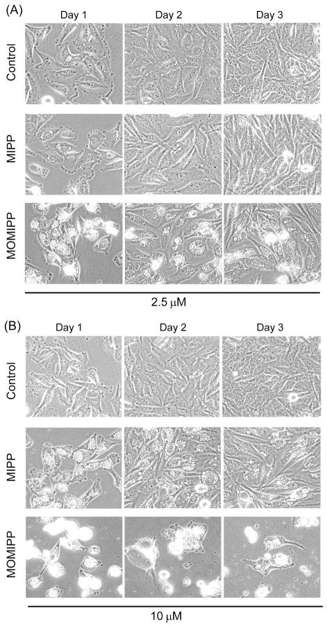
Figure 3.
Comparison of the abilities of MOMIPP and MIPP to induce the morphological hallmarks of methuosis. One day after plating, U251 GBM cells were treated with MOMIPP or MIPP at final concentrations of 2.5 μM (A) or 10 μM (B). Controls received an equivalent volume of vehicle (DMSO). Cells were observed by phase contrast microscopy on three sequential days after addition of the compounds, without changing the medium or replenishing the compounds. Methuosis is characterized by extensive accumulation of phase-lucent cytoplasmic vacuoles, with eventual cell rounding and detachment from the substratum as viability is compromised. 6,10

