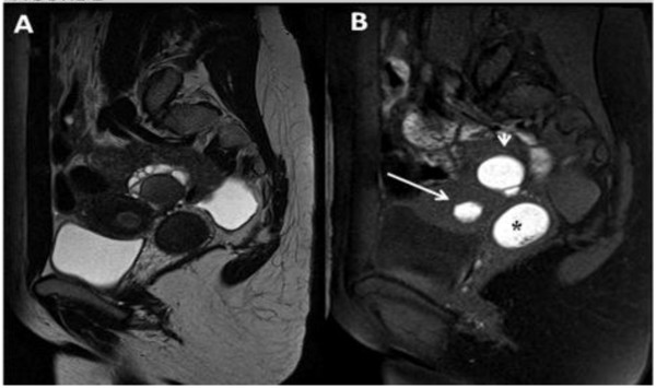
Figure 2.
A) Sagittal T2 TSE weighted image showing a mild fluid in the pouch of Douglas. (B) Sagittal T1 TSE fat-suppressed weighted image showing distention of the right endometrial cavity (long straight arrow) with high-signal-intensity material distending the right endometrial cavity and cervix (asterisk), a finding indicative of right hematometrocolpos; on the right ovary presence of an endometriosic cysts appearing hyperintense on T1 fat-saturated image (short straight arrow).

