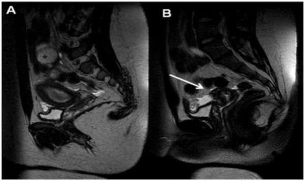
Figure 8.
A) Sagittal T2-weighted MR image shows a normal left uterine cavity communicating with a normal cervix (long straight arrow), which in turn communicates with a normal vagina. (B) Right hematosalpinx (asterisk) with a distended non-communicating horn filled with fluid moderately hyperintense on T2 sequences due to haemoglobin degradation products.

