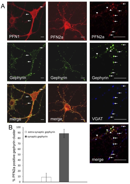
Figure 4. PFN2a is enriched in postsynaptic regions of inhibitory synapses.
(A): Confocal images of neurons with simultaneous immunostaining of PFN1 (left column), PFN2a (centre column) and gephyrin, a protein concentrated in the active zone of inhibitory postsynapses. (cf. [2]). Note that PFN2a is frequently concentrated in gephyrin clusters, while PFN1 is rarely enriched in these structures (arrows). Right column: Higher magnification of a neuron immunostained for PFN2a, gephyrin and VGAT, a marker for the inhibitory presynapse. Note that PFN2a primarily colocalises with synaptic gephyrin clusters (arrows), whereas extra-synaptic gephyrin clusters, identified by lack of VGAT staining, are mostly negative for PFN2a (arrow heads). Bar: 10 µm. (B): Quantitative analysis of the presence of PFN2a in synaptic and extra-synaptic gephyrin clusters (at least 483 extra-synaptic and 1830 synaptic gephyrin clusters per experiment, mean errors are based on 3 independent experiments, statistical analysis by unpaired t test).

