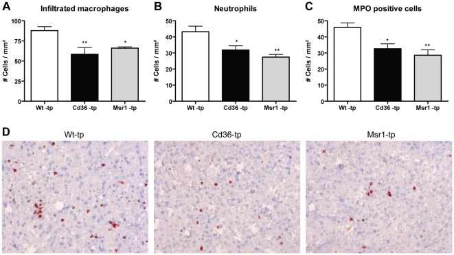
Figure 1. Parameters of hepatic inflammation.
(A–C) Liver sections were stained for infiltrated macrophages and neutrophils (Mac-1), neutrophils (NIMP) and MPO (D) Representative pictures of Mac-1 staining (×200 magnification) after 3 months of HFC feeding in Wt-tp, Cd36−/−-tp and Msr1−/−-tp mice. *Significantly different from Wt-tp group. * and ** indicate p<0.05 and 0.01, respectively.

