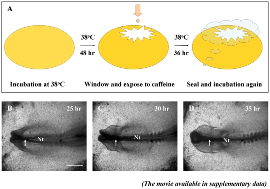
Figure 1. The strategy for administering caffeine to early chick embryo in vivo (A) and normal chick embryo neurulation (B–D).
A: Schematic drawing of caffeine introduction into early chick embryo in vivo. The fertilized egg was windowed on day 1.5, treated with caffeine, then sealed and continually incubated until the required stage. B–D: Photographed images of a normal chick embryo development, taken at the incubation 25, 30 and 35 hours. The movie version can be found in the Movie S1. The white arrows indicate the process of neural tube closure. Scale bar = 500 µm in B–D. Abbreviation: Nt, neural tube.

