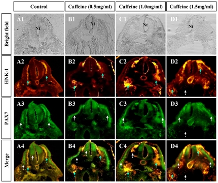
Figure 3. Caffeine induced discontinuity in cranial neural crest cells migration during delamination.
A: The control embryo, which was treated with physiological saline solution. B–D: Caffeine-treated embryos of different concentrations. A1–D1: Transverse sections of the control and caffeine-treated embryos at the cranial level. The morphology of the embryos appears normal at caffeine concentrations of under 1.5 mg/ml, above which the structure of the neural tube is damaged (D1). A2–D2: Transverse sections of whole-mount immunocytochemistry against HNK1. In the caffeine-treated embryos, the HNK1 labeled migratory neural crest cells showed a disjointed migration trajectory, as indicated by the blue dotted arrows (B2–D2). The normal continuous migration trajectory was demonstrated by the control (A2). A3–D3: Transverse sections of whole-mount immunocytochemistry for Pax7. In the caffeine-treated embryos, the Pax7 labeled neural crest cells followed a relatively normal migration trajectory (B3–C3). However, at 1.5 mg/ml concentration of caffeine, there is some structural damage as indicated by the white arrows in D3. A4–D4: Images of HNK1 (A2–D2) merged with Pax7 (A3–D3). Scale bar = 100 µm in A1–D4. Abbreviation: Nt, neural tube.

