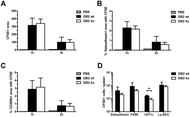
Figure 4. Sialylation of C. jejuni increases in vivo phagocytosis by splenic DC but not macrophages.
Mice were sacrificed 1 h or 3 h after i.v. injection of PBS or CFSE-labelled GB2 (wt or Cst-II mutant). Bacterial deposition in spleen sections, stained for sialoadhesin, CD209b and B220, was quantified as detailed in the Materials and Methods section. The CFSE-positive area of digital images was similar between GB2 wt and Cst-II mutant injected animals (A). No differences were observed in the CFSE-positive area within MOMA1 (B) or CD209b-positive cells (C). For each mouse, 6 pictures from 2 spleen sections were used for analysis (n = 4). Using Facs-analysis, the number of CFSE-positive metallophilic macrophages (Sialoadhesin+), red pulp macrophages (F4/80+), DC (CD11c+) and neutrophils (Ly-6G/C+) was determined per spleen (n = 3; D). Shown are means ± sd. * p<0.05; t-test with Welch's corrections.

