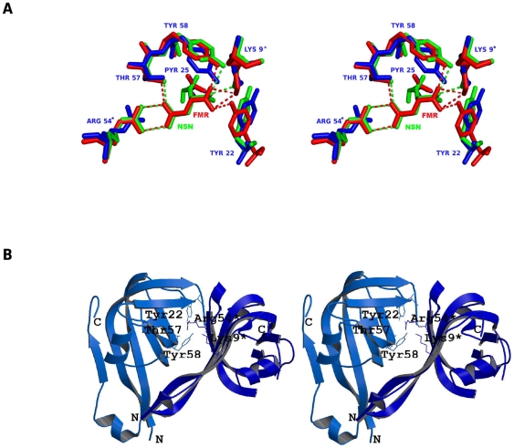
Figure 1. Conserved functional residues of ADCs that bind to substrate.
(a) Stereo view of structural superimposition of processed MtbADC (blue), processed Thermus thermophilus ADC complexed with substrate analog fumarate (red and PDB id: 2EEO) and Helicobacter pylori ADC complexed with substrate analog isoasparagine (green, PDB id: 1UHE). The conserved and interacting residues are labeled according to MtbADC and the interactions are shown as dashed lines. (b) Stereo view of the active site in the dimer interface. The figure was prepared using Molscript [36] and Raster3D [37].

