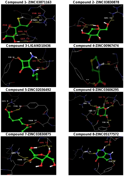
Figure 3. Binding poses of the identified eight lead molecules with MtbADC.
The binding modes of the proposed lead molecules are shown as ball and stick. Atoms colors are: H: white, C: green, N: blue, O: red and S: yellow. The interacting MtbADC residues are drawn as thin wireframe in the same color scheme and are labeled. Hydrogen bond interactions are shown as dotted yellow lines, along with the distance between donor and acceptor atoms. The binding pose of protein:lead molecule interactions were generated with the Maestro program in the Schrodinger software suite.

