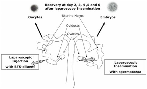
Figure 1. Schematic representation of the experimental design.
Sows were subjected to laparoscopic surgery. While one oviduct was subjected to laparoscopic insemination with (3×10⁵ spermatozoa/100 µl) spermatozoa (inseminated side), the contralateral oviduct was injected with BTS-diluent containing no sperm (non inseminated side). Oviductal and uterine horn samples as well as flushings were collected from both horns at day 2, 3, 4, 5 and 6 after laparoscopy insemination by hysterectomy. The presence of embryos at different stages of pregnancy in one horn (inseminated side), and the existence of unfertilized oocytes in the other horn (non inseminated side) were verified by careful examination of flushings under a stereomicroscope.

