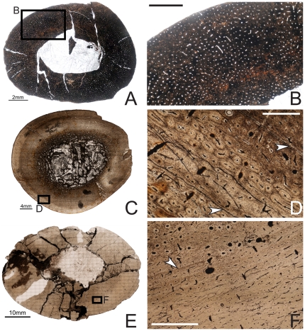
Figure 2. Osteohistology of the mid-diaphyseal humerus of Tenontosaurus in a juvenile (A, B), subadult (C, D) and adult (E, F).
A. Cross-section of OMNH 10144. This bone was invaded by bacteria before fossilization and thus much of the primary tissue is obscured. It is presented here in cross-section to illustrate vascular density and arrangement. B. Detail of A, showing general vascular patterning. The cortex is dominated by longitudinal canals arranged circumferentially. C. Cross-section of OMNH 8137. D. Detail of C, showing primary cortical tissues. The bone is woven, and most canals are longitudinal primary osteons (some anastomose circumferentially). Two LAGs (arrows) are shown. E. Cross-section of FMNH PR2261. F. Detail of E, showing mostly primary tissues of the midcortex at a transition to slower growth. Deeper in the cortex (upper left), bone is woven and osteocytes are dense and disorganized. Some secondary osteons are visible, but they do not overlap or obscure all of the primary tissues. Past the LAG (arrow), canals remain dense but decrease in diameter, bone tissue is weakly woven, and osteocytes decrease in number and become more organized. Scale bars: A = 2 mm; B, F = 1 mm; C = 4 mm; D = 0.5 mm; E = 10 mm.

