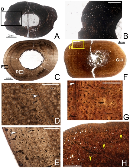
Figure 3. Osteohistology of the mid-diaphyseal ulna of Tenontosaurus in a juvenile (A, B) and two subadults (C–E, F–H).
A. Cross-section of OMNH 10144. This bone was invaded by bacteria before fossilization and thus much of the primary tissue is obscured. It is presented here in cross-section to illustrate vascular density and arrangement. B. Detail of A, showing general vascular patterning. The cortex is dominated by longitudinal canals arranged radially. C. Cross-section of OMNH 2531. D. Detail of the midcortex of C, showing primary cortical tissues. Longitudinal primary osteons are visible in primary woven bone tissue. One LAG (arrow) is shown. E. Detail of the periosteal region of C, showing the surface condition in ulnae that lack the middiaphyseal rugosity. Longitudinal primary osteons are visible in primary woven bone tissue, very similar to the tissue of the midcortex. F. Cross-section of OMNH 34191. G. Detail of the midcortex of F, showing primary cortical tissues. Midcortical tissues are similar to those shown in OMNH 2531. One LAG (arrow) is shown. H. Detail of the periosteal region of F, showing the periosteal condition in ulnae with a rugosity on the posterior surface of the bone at midshaft. The histology of this rugosity is a strongly woven, high vascularized, and very disorganized tissue that builds along a surface similar to that seen in E (yellow arrows). It grades laterally into and is capped by more typical primary bone tissue identical to that of the rest of the cortex. Scale bars: A = 2 mm; B = 1 mm; C,F = 4 mm; D,E,G = 0.5 mm; H = 2 mm.

