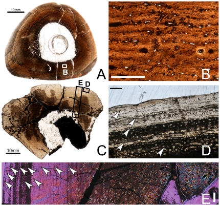
Figure 6. Osteohistology of the mid-diaphyseal femur of Tenontosaurus in a subadult (A,B) and adult (C–E).
A. Cross-section of OMNH 34784. B. Detail of A, showing the primary cortical tissues of the cortex. Longitudinal primary osteons begin to form circumferential anastomoses in the woven bone tissues of the inner and midcortex. C. Partial cross-section of FMNH PR2261. D. Detail of C, showing the tissues of the periosteal region. Longitudinal primary osteons and simple canals are not as dense as in the midcortex and anastomose less frequently moving periosteally. Tissue is lamellar. Five LAGs (arrows) are shown. E. Detail of a radial transect through the cortex of C. Image taken through waveplate polarizing filters (crossed Nicols). Dense secondary remodeling is visible into the midcortex and zones of decreasing width are visible. Ten LAGs (arrows) are shown. Scale bars: A,C = 5 mm; B,D = 0.5 mm; E = 1 mm.

