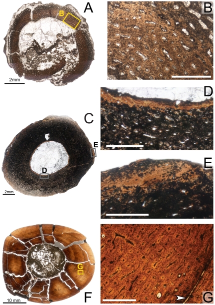
Figure 7. Osteohistology of the mid-diaphyseal tibia of Tenontosaurus in a perinate (A,B), juvenile (C,D), and subadult (F,G).
A. Cross-section of MOR 788. B. Detail of the primary cortical tissues of A. Longitudinal simple canals and primary osteons have wide diameters compared to those of older ontogenetic age. Bone tissue is woven-fibered. C. Cross-section of OMNH 10144. This bone was invaded by bacteria before fossilization and thus much of the primary tissue is obscured. It is presented here in cross-section to illustrate vascular density and arrangement. D. Detail of the endosteal region of C, showing lamellar tissues (to right of image). Canals are narrower in diameter compared to the perinate (B). E. Detail of the periosteal region of C showing primary cortical tissues. The longitudinal primary osteons are surrounded by woven bone tissue. F. Cross-section of OMNH 63525. G. Detail of the midcortex of F. Longitudinal primary osteons run through woven bone tissue and show short circumferential anastomoses. One LAG (arrow) is shown. Scale bars: A,C = 2 mm; B,D,E,G = 0.5 mm; F = 5 mm.

