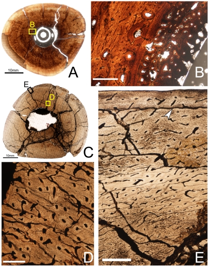
Figure 8. Osteohistology of the mid-diaphyseal tibia of Tenontosaurus in a subadult (A,B) and an adult (C,D,E).
A. Cross-section of OMNH 34784. B. Detail of the inner cortex of A showing the endosteal surface and medullary bone (egg-laying) tissue. The primary cortical tissue (left side) consists of woven bone vascularized by longitudinal primary osteons connected by moderately long circumferential anastomoses. This tissue is beginning to undergo secondary remodeling. Endosteal lamellae separate the cortical bone from the medullary bone tissue, which radiates inward into the medullary cavity. C. Cross-section of FMNH PR 2261. This specimen was treated with oil before photography to increase light penetration, but this reduces the appearance of some thin, mineralized structures (LAGs, cement lines). D. Detail of C, showing histology of the inner cortex. Secondary osteons are abundant and obscure much of the primary cortical tissue. E. Detail of C, showing the outer cortex. Osteocytes are dense throughout the cortex, despite the transition to parallel-fibered bone in this region. Canals of the outermost cortex anastomose less frequently compared to the inner cortex. Scale bars: A = 5 mm; B,D,E = 0.5 mm; C = 10 mm.

