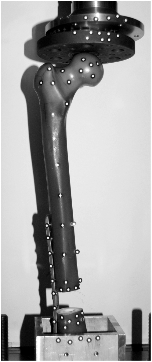
Figure 5. Experimental test setup.
Posterior view of the test arrangement with a composite left femur mounted in the universal testing machine (Zwick/Roell). Segmental defect is bridged with an osteosynthesis system on the lateral (outer) side and fixed with seven titanium screws. Distal end of the femur is embedded in a metallic socket, filled with casting resin. 57 optical markers were attached onto the femur, socket and the testing machine to calculate their displacement during loading.

