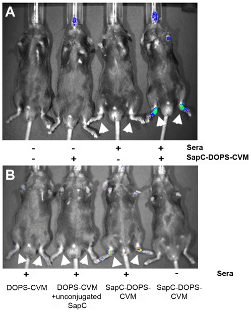
Figure 1. SapC-DOPS-CVM localizes to arthritic joints and is visible by live fluorometric imaging.
Male C57Bl/6 mice three months of age were given 150 µl of sera i.p. as indicated. Seven days after sera injection, mice were imaged by IVIS® (A) five hours following SapC-DOPS-CVM i.v. injection as indicated (- is PBS); (B) two hours following i.v. injection of DOPS-CVM, DOPS-CVM plus uncoupled SapC, or SapC-DOPS-CVM as indicated. Arrows indicate macroscopically swollen paws.

