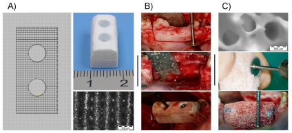
Figure 7. Implantation in pig maxillary defects: materials and surgery.
A) Robocast ceramic design, macroscopical appearance of scaffold and microscopical detail. B) Images of surgery procedure, implantation of a Robocast sample and fixation of it with screws. C) Microscopical image of a Bio-Oss® sample, detail of Bio-Oss® sample preparation procedure and Bio-Oss® implanted and fixated with surgical glue (see blue glue between sample and surrounding bone).

